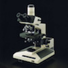
Brightfield
We have two Olympus BH-2 brightfield microscopes. Both are equipped with color cameras, plan Apo lenses to 100x, epifluorescence capability using a wide variety of custom and standard filter cubes and differential interference contrast (Nomarski) optics.
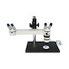
Double-Headed Dissecting
This dissecting microscope is ideal for instruction purposes and projects which require the expertise of more than just one person. Its large base makes it ideal for using on bigger samples, which would be otherwise precarious on smaller microscopes.
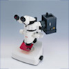
Fluorescent Dissecting
The Leica MZ FLIII fluorescence stereomicroscope offers not only a 3D image but also, compared with a classical microscope at any given magnification, a larger panoramic field of view, more intense fluorescence, and longer working distances.
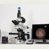
Hyperspectral
The CytoViva Hyperspectral Imaging system allows for spectral characterization and spectral mapping of nanoscale samples. The technology can also be used with a wide range of other types of samples, from micro to macro in scale, and for a number of applications. Importantly, it allows visualization of unstained nanoscale objects in tissues and cells.
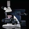
Laser Capture
Whether for cancer research, pathology, proteomics, genomics, gene expression profiling or neuroscience, the new, state-of-the-art LMD 6000 laser microdissection system from Leica Microsystems captures tissues and cells easily and quickly. Users appreciate the intuitive touch screen interface and the speed of this system.
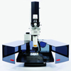
Laser Scanning Confocal
Leica TCS LSI is the first super zoom 3D-confocal, offering high resolution plus large field of view. The new LargeScale Imaging (LSI) platform provides generous workspace and adapts perfectly to the experiment needs of native specimen analysis, particularly larger specimens. With four laser excitation and tunable emission offers flexibility for multi-color fluorescence staining.