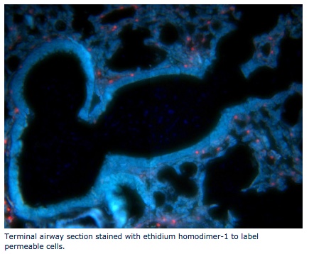
 The Cellular and Molecular Imaging (CAMI) Core provides research quality standard light and fluorescent microscopes and equipment for histology, as well as sample preparation capability in one location based on an approved recharge rate.
For new user inquiries and onboarding, please contact the CAMI Core Admin at camicore-request@ucdavis.edu.
CAMI's Areas of Expertise
- Laser capture and confocal microscopy
- Basic microscopy including fluorescence at low and high magnification
- Immunohistochemistry on sections and whole mount tissue
- Histology of tissues and cells
- Custom embedding (resin, paraffin, frozen, small or difficult samples)
- Semi- thin resin sectioning
- qRT-PCR and RNA extraction of small samples
- Teaching users who are unfamiliar with microscopy, histology, fluorescence and imaging