Overview
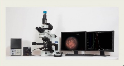
The CytoViva Hyperspectral Microscope Imaging system allows for spectral characterization and spectral mapping of nanoscale samples. Each pixel of a hyperspectral image provides the complete reflectance spectral response of that pixel’s spatial area within the visible and near-infrared (VNIR) or short-wavelength infrared (SWIR) spectral range. This enables nondestructive spectral measurements of nanoscale elements in the full spatial context of the sample image. For example, at 100x magnification, a hyperspectral image may contain as many as 700,000 pixels, each as small as 128nm each. The hyperspectral imaging technology supports a wide range of other types of samples, from micro to macro in scale, and applications.
Key Features
- Easy to use
- High spectral resolution
- Can be used with nanoscale samples
- Certain biological materials, such as bacteria, can be optically observed, spectrally characterized, and mapped in tissue
- Can be used to spectrally unmix stains, such as H and E or Alcian Blue PAS
- No special fluorescent markers required.
Recharge Rate
$33/hour for UC Davis users, $43/hour for non-UC Davis users